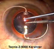
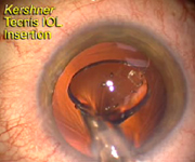
Figure 1-Kershner.
The Tecnis IOL is a three-piece silicone lens with a modified prolate
anterior surface.
|
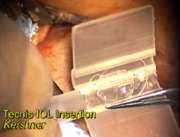
The Tecnis™ modified prolate anterior surface IOL now has an injector system.
|
|
|
Figure 1-Kershner.
The Tecnis IOL is a three-piece silicone lens with a modified prolate
anterior surface. |
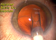
Figure 2-Kershner. The Tecnis IOL is loaded into the cartridge with the haptics positioned at the 3 o’clock and 8 o’clock positions. |
|
|
|
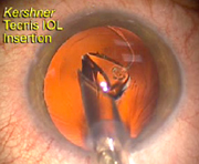
Figure 3-Kershner. The Injector cartridge is inserted bevel down. The Tecnis IOL unloads in a controlled fashion into the capsular bag with the leading haptic positioned down and to the left of the cartridge tip. |
Figure
4-Kershner. The injector and cartridge
is rotated 90º counterclockwise as the optic gently unfolds.. |
|
|
|
Figure 5-Kershner. The lens optic is delivered. |
| . |
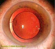
Figure
6-Kershner. The IOL need not be repositioned,
as it self-centers when the OVD is completely removed. |
|
by Robert M. Kershner, M.D., M.S., F.A.C.S. ©2004. All rights reserved.
Special to Ocular Surgery News
Many surgeons are now familiar with the characteristics of the Tecnis Z-9001 modified prolate anterior surface IOL, designed to neutralize the eye’s spherical aberration. But until recently, the IOL was at a disadvantage because of the absence of a reliable injection system that would allow surgeons to implant the IOL through a small incision.
Numerous studies have demonstrated the ability of the Tecnis to improve visual acuity and to improve contrast perception, especially at night and at night when glare is present. The Food and Drug Administration has allowed labeling designating that the lens improves functional vision and safety.
The acquisition this past summer of parts of Pfizer’s ophthalmic surgical line, including the Tecnis brand, has created an opportunity for AMO to adapt its well-known IOL injection systems to this lens style, which requires special attention with implantation.
As a surgeon who has studied the unique characteristics of this new-technology IOL, I would like to share my extensive experience with implanting the Tecnis IOL under a variety of circumstances.
Adapting to the new technology
The AMO Silver Series T injection system has now been validated for the Tecnis IOL. Combined with the Silver Series II cartridge, the marriage is complete. Surgeons have a readily available and easy-to-use system to incorporate the Tecnis technology into their armamentarium of IOLs. Because this IOL is not your average spherical implant, the unique characteristics of the modified prolate anterior surface and stiff polyvinylidene (PVDF) haptics create special requirements when loading and unloading the IOL into an injector and into the capsular bag.
The Tecnis IOL is a posterior chamber, three-piece, foldable IOL with a CapC design for posterior capsular bag fixation . The Z-9000 with a 12-mm overall length and its counterpart, the Z-9001 with a 13-mm overall length, are made of a next-generation silicone, a UV-absorbing polysiloxane. The optic is biconvex, with a patented modified prolate anterior surface and a square optic edge with an index of refraction of 1.46.
The two clear haptics, made from polyvinylidine fluoride, are known for their strength, but remain somewhat stiff during implantation. The haptics come off the IOL optic at 90° takeoffs and are angulated 6°. These characteristics of the 911A platform from which they are derived are very important in assuring proper centration as well as avoiding tilting of the IOL within the capsular bag, maximizing the potential for the optic to deliver its full benefit. Unfortunately, they are the same culprits that can make the loading, folding and unfolding of the lens problematic.
Some surgeons, in their early experience with this IOL, know what can happen when attention is not paid to the loading process. An inadvertent haptic capture in the nose cone of the cartridge by the injector plunger can crimp or bend a haptic, or worse yet, cause a complete destaking, taking a piece of the IOL optic with it. Fortunately, this can be completely avoided when the proper loading procedure is strictly followed.
The AMO Unfolder Silver Series T Injector reusable handle and Silver Series II disposable single-use cartridge are easy to use and similar to many presently in use. I have found that I can easily implant the Tecnis IOL through a sub 3-mm incision and routinely do not need to enlarge my standard 2.4-mm clear corneal incision.
When you first start loading this lens, it is best if the following procedure is done under the operating microscope. Once comfortable with the method, the lens can be loaded on the side table without magnification. I feel that demonstrating the technique for your scrub assistant by using a video monitor or by viewing through the operating microscope facilitates the learning process.
Step-by-step tips to maximize success
First, remove the cartridge from its sterile packaging and place it on the table so the nose cone faces to the left (at the 9 o’clock position). You do not need to open the cartridge completely; in fact, a slight bend to the hinge works best when positioning the IOL optic under the wings. It is easier to load this lens with the cartridge on a table for stability, rather than trying to hand hold the cartridge and the lens at the same time.
I suggest using a very small ribbon of an ophthalmic viscosurgical device (OVD) such as Healon or Healon 5 (sodium hyaluronate, AMO) on the length of the hinge and sparingly into the troughs of the cartridge. Avoid using dispersive agents such as methylcellulose, which can result in inconsistent IOL delivery and leave an undesirable coating on the optic.
When using Healon 5, which is an excellent lubricant, recognize that it is quite viscous and can actually compete with the IOL for space, so less is more; use it very sparingly to get the same effect as with the less viscous agents. If you get a little too generous with your OVD, it can work against you. There is nothing worse than an IOL covered with an OVD if you don’t get it positioned under the cartridge wings on the first try. (Think: greased pig competition.)
Remove the IOL container from its sterile package and place it flat on the table. Press on the cover and remove it. Using blunt forceps, gently slide one of the blades into the groove overlying the right-side haptic and lift out the IOL. (I prefer the modified prolate IOL lens-holding forceps made for me by Rhein Medical, Tampa, Fla. It is specially designed to hold a nonsymmetric optic without slipping or damaging it.)
With the cartridge tip pointed to the surgeon’s left, and 12 o’clock being where the tip is, load the IOL approximately 0.5 mm to 1.0 mm behind the IOL diagram shown on the cartridge wing. Position the leading haptic anchor at the 3 o’clock position and the trailing haptic anchor at the 8 o’clock position. Loading the IOL too far from the nose cone may not allow position of the leading haptic. Loading it too close may prevent the trailing haptic from remaining outside of the folded cartridge.
Before letting go of the lens, use the forceps blades to press the optic under the edges of the two wings of the cartridge. (With a little practice this goes pretty easily, as your first attempt to load the lens is usually your best shot.)
With the forceps blades removed and slightly opened, position them flat onto the surface of the lens optic to move the IOL back and forth along the edge of hinge. This assures a tactile and visual check that the optics are secured under the grooves of the cartridge wings. If they are not, reposition the lens to make sure the IOL optic is captured under the cartridge wing edges. The resilience of the optic material and the highly polished surface of the forceps blades reduce the chance of inadvertent damage to the optic.
Next, while holding the optic into position to assure that it does not pop out of the grooves, gently fold the cartridge wings. The IOL optic should have a concave configuration and be well secured under the grooves of the cartridge wings.
Manually position the leading haptic using the closed forceps tips under direct visualization so that it is directed into the cartridge nose cone and complete the closure of the cartridge.
Visually inspect that the trailing haptic remains out of the cartridge. If the trailing haptic is trapped under the wing edge or bends toward the optic, give it a gentle sweep with the forceps blade. You need not reopen the cartridge to do this.
Load the silicone tip onto the injector plunger.
Grasp the injector making sure the plunger is fully retracted. Without allowing the cartridge to open, slide the tip and nose cone into the slot of the injector.
Immediate insertion is best. Allowing the loaded injector to dry out for several minutes can encourage lens or optic capture and a torn IOL upon delivery.
I prefer not to fill the capsular bag with an OVD, but rather place a small bolus of Healon 5 into the center of the capsular opening. In this manner the unfolding of the IOL can occur unimpeded under the bolus of Healon 5 and drop directly into the capsular bag. The bolus of Healon 5 acts to push the IOL into position and, by remaining on top of the lens optic, protects the corneal endothelium while facilitating the OVD removal at the end of the case. This avoids the need to rock and roll, or tilt the IOL in order to remove the OVD trapped behind the optic.
Position the injector tip bevel down and pass it through the incision such that the tip overlies the center of the capsular opening.
Advance the plunger while directly visualizing the passage of the IOL and haptics down the injector cartridge. Do not back up the plunger while advancing the lens as this may override the optic or haptic causing an optic tear.
As the IOL unfolds, the leading haptic will position itself down and to the left of the injector tip. Slightly rotate the barrel of the injector counterclockwise 90° to facilitate the remaining unfolding process.
Once the IOL has unfolded, and the trailing haptic is freed from the injector barrel (and not before) rotate the cartridge barrel to point the tip bevel down. Retract the plunger making sure it does not capture the trailing haptic as it does so, and use it as a positioning tool to engage and place the trailing haptic into the capsular bag. Alternatively, if the trailing haptic fails to release, use a forceps in the nondominant hand to remove the haptic and manually position it into the bag. This is the most common stumbling block for surgeons inexperienced with this lens insertion. A little patience here pays off.
It is not necessary to position the haptics in any preferred orientation, so dialing the lens or moving it after it is implanted is best avoided once the lens is in place.
Remember all good practitioners clean up after themselves. Never leave an OVD in the eye at the conclusion of surgery anymore than you would leave an IOL forceps. Visualize the complete irrigation and aspiration of any remaining OVD from in front of, adjacent to or behind the lens. A slight malpositioning of the Tecnis optic is the telltale sign that there is trapped OVD. Once the remaining OVD is removed, this IOL will self center without help.
Adopting the technology of the Tecnis modified prolate IOL into your practice will more than reward the perseverant surgeon with better cataract surgery outcomes and happier patients. The technology is here today and it is easier than ever before to adopt into your practice.
For Your Information
Robert M. Kershner, M.D., M..S, F.A.C.S., can be reached at Eye Laser Consulting, Boston, Massachusetts USA. www.EyeLaserConsulting.com/PhysicianResource.htm. Dr. Kershner has no financial or proprietary interest in any product or company mentioned. Tecnis™ and Healon™ are trademarks of AMO, Inc. Santa Ana, CA USA. For more information on the Tecnis IOL the author refers you to: www.TecnisIOL.com.
Article and Images: ©2004. R.M. Kershner , M.D., M.S., F.A.C.S., Eye Laser Consulting, Boston, Massachusetts USA