Clear Corneal Cataract Surgery and the Correction of Myopia, Hyperopia and Astigmatism

Robert M. Kershner, MD, FACS Copyright.

Eye Laser Center, Suite 303, 1925 West Orange Grove Road, Tucson, Arizona USA 85704-1152, Phone (520) 797-2020, e-mail: kershner@EyeLaserCenter.com

26 Pages

Abstract

Purpose: Advances in cataract removal using topical anesthesia through a clear corneal microincision have created a new opportunity to fully correct refractive errors at the time of cataract surgery. This study was undertaken to assess the effectiveness of keratolenticuloplasty (KLP), the simultaneous modification of the cornea at cataract removal to create emmetropia with lens implantation.

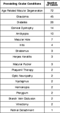
Methods: Data were analyzed for 690 consecutive cataract procedures performed between March 1993 and March 1995 with followup of 12 to 24 months. Each patient underwent cataract removal with topical anesthesia, clear corneal incision fashioned as an arcuate keratotomy to correct preexisting astigmatism, intercapsular phacoemulsification, and microinjection of a single-piece elastic intraocular lens (IOL) into the capsular bag to correct spherical error.

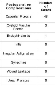
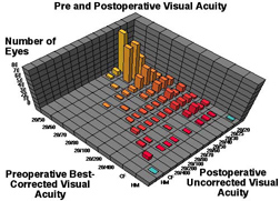
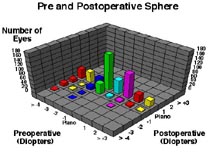
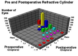
Results: Preoperative, best corrected, visual acuity was less than 20/50 in all patients; 58% were myopic, 32% were hyperopic, and 57% had astigmatism of greater than 1 diopter (D). Postoperatively, spectacle independence was achieved with uncorrected vision of 20/40 or better in 87% of eyes. The sphere was fully corrected in 78%, within 1 D in 17%, and within 2 D in 5%. No patients were overcorrected. The cylinder was fully corrected in 72%, within 1 D in 26% and within 2 D in 2%. Of those with residual astigmatism, there was no significant postoperative shift in cylinder axis. There were no sight threatening complications. All patients were able to resume normal unrestricted activities within twenty-four hours of undergoing the procedure.

Conclusion: The KLP technique can correct spherical and astigmatic refractive errors creating spectacle-free individuals following clear corneal cataract surgery.


Clear Corneal Cataract Surgery-Kershner

Page 2

Introduction

Clear vision without the need for eyeglasses or contact lenses has been one of mankind's goals. Advances in the technique of modern cataract removal has created a new opportunity to provide clear, uncorrected vision for patients who undergo cataract surgery. The introduction of phacoemulsification for cataract removal1 and its increasing acceptance, 2 has made it possible for surgeons to take full advantage of the small incision that this procedure requires. Smaller incisions have reduced the need for suturing and its attendant induction of astigmatism.3-6 With smaller incisions of 3.0 mm or less, the incision can be moved onto the cornea precluding the need for conjunctival dissection and cautery.7

The introduction of topical anesthesia as a substitute for retrobulbar anesthetic injection for cataract surgery,8-10 changed the approach to the cataract incision.11,12 With topical anesthesia, the patient can fixate during the operative procedure, making it possible to apply refractive surgical techniques at the time of cataract surgery.13,14 These smaller incisions can induce less astigmatism.3,6,15 Several surgeons have advocated placing the corneal incision temporally,15-17 or on the steep axis,18 to reduce preexisting astigmatism. By fashioning the clear corneal cataract incision as an arcuate keratotomy, preexisting astigmatism can be simultaneously corrected at the time of cataract surgery.13,14

The increasing acceptance of newer foldable and injectable IOLs in small incision surgery19,20 has made it possible to reduce incision size to less than 3.0 mm (microincision). The preexisting spherical myopia or hyperopia can be corrected with judicious selection of a one-piece injectable IOL. Postoperative bandaging previously required following cataract surgery can be eliminated enabling faster visual recovery


Clear Corneal Cataract Surgery-Kershner

Page 3

and a more rapid return to normal activities for the patient.9

These recent technological advances can improve uncorrected vision and reduce dependence upon spectacles following cataract surgery. I have named the cataract procedure, which remodels the cornea to correct astigmatism and corrects the spherical refractive error with intraocular lens implantation, keratolenticuloplasty or KLP.13,14

This study was undertaken to assess the visual and refractive outcome of simultaneously correcting myopia, hyperopia, and astigmatism with cataract surgery. Topical anesthesia, clear corneal cataract incision positioned on the steep axis and fashioned as an arcuate keratotomy to correct astigmatism, capsulorhexis, hydrodissection, intercapsular phacoemulsification, and implantation of an injectable IOL through the same incision into the capsular bag to correct the sphere, are integrated into a refractive cataract procedure.


Clear Corneal Cataract Surgery-Kershner

Page 4

Methods

Selection of patients: All cataract procedures were performed by one surgeon (RMK), using the following protocol. Data were compiled from the preoperative examinations and the last examination of record for each patient during the study period of March 1993 until March 1995. Excluded during this period were 93 cases which involved combined procedures (26), preexisting conditions which precluded corneal incisions (43), or deviations from the protocol which prevented implantation of the one-piece capsular lens, such as capsular rupture (18). The intraocular lens was removed at the time of surgery in 6 patients due to torn capsules or failure to properly center and those cases were not enrolled. A total of 690 consecutive procedures are the basis for the data reported in this study.

Surgical protocol: All patients enrolled underwent a comprehensive ophthalmological examination. Best corrected visual acuity was determined using a Snellen acuity chart. Cycloplegia was obtained using one drop of 1% cyclopentolate hydrochloride (Cyclogyl, Alcon, Ft. Worth, Texas USA) and retinoscopic refraction performed with an Optec 360 retinoscope thirty minutes following instillation of drops. The refraction was refined with a Reichert phoropter. Applanation tonometry, slit lamp and funduscopic examination were performed.

Those patients with a visually significant cataract and subjective complaints of decreased vision attributed to the cataract were enrolled in the study. A discussion of the risks, complications and alternatives to cataract surgery and the simultaneous correction of refractive error was undertaken with each patient. An informed consent which met the requirements of the Institutional Review Board for the facility was obtained in all cases.


Clear Corneal Cataract Surgery-Kershner

Page 5

Preoperative candidates underwent A-scan ultrasonic biometry with a Sonomed ultrasonography unit. Keratometry was performed with a Bausch and Lomb keratometer. Calculations of lens power were computed using three different programs; Binkhorst I, Regression, and Colen. The lens was selected to achieve a projected postoperative refractive result of -0.25 D to -0.50 D. An A-constant of 118.5 was employed. No alteration was made in lens power if combined astigmatic keratotomy was to be performed.

The intraocular lens utilized in this study was the STAAR AA4203, a one-piece injectable silastic lens (STAAR Surgical, Monrovia, California, USA).

All patients with astigmatic errors in excess of 1.00 D, as determined by refraction, received corneal topographic analysis performed with the Tomey TMS-1 system. A surgical plan was created using the proper IOL power and a map of the arcuate corneal incision(s) on the axis of steepest plus cylinder, (as determined by retinoscopic refraction and confirmed by topographic analysis). Where there was a discrepancy between the refraction and the topographic map for cylinder axis, preference was given to the topographic analysis using the simulated keratometry readings and the qualitative appearance of the astigmatic pattern. The postoperative goal was to undercorrect with-the-rule astigmatism and overcorrect against-the-rule astigmatism by 0.25 D to 0.50 D.

Surgical procedure: The operative eye was prepared for surgery with 1 drop of 1% cyclopentolate hydrochloride, and 2.5% phenylephrine hydrochloride (Cyclomydril, Alcon, Ft. Worth, Texas USA) starting 20 minutes prior to surgery with drops repeated once every five minutes for a total of three drops. The technique of topical anesthesia has been previously published.9,10 Topical 0.5% tetracaine was instilled five minutes


Clear Corneal Cataract Surgery-Kershner

Page 6

prior to surgery, and repeated at the start of the prep. Topical povidone iodine solution (Betadine 5%, Escalon, Lakewood, NJ USA) was instilled 10 minutes prior to surgery into the operative eye. The eye and eyelid skin were prepared and draped. The corneal maps were brought into the operating room and consulted when creating the corneal incisions. The proper optical zone for the arcuate incisions based on preoperative astigmatism was selected using the Kershner nomograms.13 The KLP procedure followed the previously published technique13,14 with the following modifications:

Patients without preexisting astigmatism (73), received a single, parallel to the iris plane (planar), 2.5 mm stab incision on the oblique (120o OD, 60o OS) or temporal axis (1800) just anterior to the vascular arcade on clear cornea (Figure 1a). All eyes with 1.00 D or less of preexisting astigmatism (398) had a single arcuate 2.5mm incision on the steepest axis just anterior to the vascular arcade at the 11 mm optical zone. Eyes with 1.00 D to 2.00 D of cylinder (144) had a single arcuate 3.0 mm incision on the steepest axis at the 10mm to 11mm optical zone (Figure 1b). An additional arcuate keratotomy incision (75 eyes) was placed on the opposite end of the same meridian for preexisting astigmatism of greater than 2.00 D (Figure 1c), with smaller optical zones utilized for greater astigmatic corrections for this incision as determined by the Kershner nomograms. The patient was instructed to fixate on the light of the operating microscope, and the appropriate optical zone marker (Kershner Arcuate Keratotomy Marker - Rhein Medical, Tampa, Florida USA) was pressed onto the surface of the cornea creating two arcuate marks. Ultrasonic pachymetry was measured using a Sonogauge ultrasonic pachymeter at the two incision sites. A trifaceted, triple edge diamond keratome (Kershner Arcuate Keratotomy Diamond-Diamatrix, The Woodlands, Texas USA) was calibrated under a micronscope, and set


Clear Corneal Cataract Surgery-Kershner

Page 7

to 100% of the pachymetry measurement. The globe was fixated with a Livernois fixator (Rhein Medical, Tampa Florida), and the single or double arcuate marks were incised the appropriate length, at the proper optical zone and axis.

The arcuate keratotomy at the surgical limbus closest to the surgeon (10-11mm optical zone), was utilized for the subsequent cataract procedure. A 2.85 mm trifaceted diamond keratome (Kershner Corneotome-Diamatrix, The Woodlands, Texas USA) was placed at the base of the arcuate incision to enter the anterior chamber parallel to the iris plane and perpendicular to the arcuate keratotomy at approximately 85% of corneal thickness. The techniques of capsulorhexis, 21,22 hydrodissection,23 intercapsular phacoemulsification,24 and intercapsular implantation of the injectable intraocular lens,13,15,25 were then followed in order. Viscoelastic was used for the capsulorhexis and filling of the capsular bag for IOL implantation (Healon-Pharmacia, Inc. Dublin, Ohio USA). Phacoemulsification was performed with the Alcon 10,000 master phacoemulsification unit using linear surgeon control with a 30 degree phacoemulsification tip and custom-fitting teflon sleeve at maximum phaco powers of 30% - 60%. A STAAR microinserter with disposable cartridge was used to insert the lens through the limbal, arcuate keratotomy incision into the capsular bag. All viscoelastic was removed with irrigation and aspiration. The incision was not sutured. No intracameral antibiotics or miotics were used. The total operative time for the procedure averaged four to six minutes. The eye was not patched at the conclusion of the procedure.

Postoperative Procedure: Patients were started on topical tobramycin-dexamethasone solution (Tobredex-Alcon, Ft. Worth, Texas USA) the day of the procedure, one drop into the operative eye four times a day and continued for ten days.


Clear Corneal Cataract Surgery-Kershner

Page 8

No other topical or oral medications were prescribed. Patients were instructed to continue any previously prescribed topical glaucoma medications. Patients were asked to refrain from eye rubbing and swimming during the first two postoperative weeks, otherwise their activities were not restricted.

Patients were seen on the first postoperative day, at two weeks, one month, three months, six months and each year thereafter, unless circumstances necessitated more frequent visits. All patients underwent measurement of uncorrected visual acuity, slit lamp examination, and applanation tonometry on each postoperative visit. Refraction was performed on the last postoperative examination during the study period. The data were collected from the last visit of each patient during the study period.

All patients were surveyed as to satisfaction with their vision at one year. They were asked to score their level of satisfaction as very satisfied, satisfied or not satisfied. They were further asked if they used eyeglasses for distance only, distance and reading, reading only or not at all.

Results

A total of 690 surgical procedures which adhered to the above protocol were included in the study (690 eyes of 587 patients). Followup was completed for 538 patients at two years, and 49 patients at one year. Data were included for 15 patients who died and 23 patients who were lost to follow-up during the course of the study, but completed the one year exam.

The average age of the patients in the study was 72.6 years (range 41 to 102 years). Male to female ratio was 1:1.4 . The ratio of right eyes to left eyes were 1.09:1.0.

The distribution of preoperative best-corrected and postoperative uncorrected visual acuities is shown in Figure 2. Preoperatively, 63% of eyes had best corrected visual acuities of 20/50 to 20/70 with the remainder (37%) being 20/80 or worse.


Clear Corneal Cataract Surgery-Kershner

Page 9

Uncorrected visual acuity improved to 20/40 or better in 87% of eyes following surgery. Uncorrected vision of only 5% of eyes were in the range of 20/50 to 20/70 following surgery.

The pre and postoperative spherical refractive error is shown in Figure 3. Preoperatively, 58% of eyes were myopic, 10% had no refractive error (<0.25 D), and 32% were hyperopic. Spherical error was reduced to plano in 78% of cases, -1 D or less in 17%, and -2 D or less in 5% postoperatively.

The pre and postoperative refractive cylinder is shown in Figure 4. Preoperatively, 10% of eyes had no astigmatism (<0.25 D), 33% had +0.5 D, 24% had +1 D, 21% had +1.5, 7% had +2 D, 2% had +2.5 D and 3% had +3 D or more. Of those eyes with preexisting astigmatic error, corneal astigmatism was eliminated in 72% following the surgery, reduced to +0.5 D in 14%, +1 D in 12%, and +1.5 D in 2%. Preoperatively those eyes that required a single, arcuate keratotomy, averaged +1.44 D (±0.69 S.D.) of corneal astigmatism, with a reduction of postoperative cylinder power to +0.78 D (±0.32 S.D.). In those eyes that required two arcuate keratotomy incisions to correct greater than 2 D of preexisting astigmatism, the average preoperative cylinder was +2.56 D (±0.82 S.D.) with a reduction of postoperative cylinder power to +0.68 D (±0.57 S.D.), demonstrating the powerful effect of the additional incision.

The effect of arcuate keratotomy on shifting the axis of the astigmatism postoperatively was analyzed by the method of Retzlaff, Paden and Ferrell.26 Of the 193 eyes (28%) that had residual astigmatism following the procedure, only 3% had a shift in axis of greater than 15o. Vector analysis of the effect of the arcuate keratotomy procedure on the magnitude and direction of the preoperative astigmatism is depicted in Figure 5. The 148 eyes with more than +0.75 D of astigmatism following the


Clear Corneal Cataract Surgery-Kershner

Page 10

procedure are shown by location of the astigmatism, for each axis group and broken out by those requiring a single incision arcuate keratotomy or two arcuate incisions.

For those eyes that received a single keratotomy incision, the against-the-rule (ATR) group (n=91), had preoperative cylinders averaging +1.29D (± 0.36 standard deviation, S.D.). The induced astigmatism from the surgery is illustrated in the center circle by a with-the-rule (WTR) shift of +1.04 D, (± 0.41 S.D.) with residual astigmatism of +0.74 D (± 0.35 S.D.) in the ATR direction. The WTR group (n=13) averaged +1.21 D (±0.32 S.D.) of preoperative cylinder, +0.96 D (± 0.65 S.D.) of surgically-induced cylinder and a resulting cylinder of +0.75 D (±0.40 S.D.). The left oblique (LO) group (n=9) preoperatively averaged +1.08D, (± 0.33 S.D.) with surgically induced cylinder in the right oblique (RO) direction of +0.73 D, (±0.57 S.D.) for a resulting cylinder of +0.69 D (±0.30 S.D.). The RO group (n=4) averaged +1.13D (± 0.43 S.D.) preoperatively, with surgically induced cylinder in the LO direction and to a lesser extent in the ATR direction (+0.74D ±0.38) with a net result of +0.63D (± 0.25 S.D.).

For those eyes that received two keratotomy incisions, the ATR group (n=25), had preoperative cylinders averaging +2.68 D (± 0.88 S.D.). The induced astigmatism from the surgery equalled + 2.19 D, (±1.02 S.D.). The ATR group had their astigmatism reduced by 60% to +1.06 D (± 0.47 S.D.) in the ATR direction. The WTR group (n=5) averaged +3.05 D (± 0.95 S.D.) of preoperative cylinder, +2.28 D (±1.35 S.D.) of surgically-induced cylinder in the ATR direction with a resulting cylinder of +1.2 D (±0.44 S.D.) in the WTR direction. The LO group (n=1) had +3 D (±0 S. D.) preoperatively, with surgically induced cylinder in the RO direction of+2.05 D (±0 S. D.), for a residual astigmatism of +0.75 D (±0 S. D.) in the LO position. There were


Clear Corneal Cataract Surgery-Kershner

Page 11

no cases in the RO group. In all cases of residual astigmatism, the procedure more than halved the preexisting cylinder, without reversing it. The results of pre and postoperative topographic analysis have been previously published13 and will not be addressed in this report.

The list of preexisting ocular conditions in this series are shown in Table 1. Of the remaining 11% of eyes that did not achieve uncorrected vision of 20/40 or better, all but 26 cases had an improvement of two lines or more of vision from their preoperative levels. Those cases that lost more than one line of postoperative best-corrected visual acuity had preexisting ocular disease as follows: 2 retinal buckling procedures for retinal detachment with preexisting macular cellophane, 6 refractive amblyopes, 2 optic neuritis, 4 age-related macular degeneration (ARMD), 4 macular holes, 1 branch vein occlusion, 1 prior vitrectomy with macular traction, 1 cystoid macular edema (CME), 2 corneal dystrophy, and 3 prior trabeculectomy for chronic open angle glaucoma.

Postoperative complications are listed in Table 2. CME occurred in 3 eyes, as defined by loss of two or more lines of visual acuity and documented by intravenous fluorescein angiography. All three cases of CME resolved with treatment. Capsular fibrosis occurred in 48 eyes. YAG laser capsulotomy was performed in those cases where visual acuity was impaired to less than 20/40. The earliest procedure was performed at 3 months and the latest at 2 years (6.9%). There were no post YAG complications. There were no cases of irregular astigmatism, postoperative synechiae, iritis, wound leakage or uveal prolapse.

The most serious complication of surgery was one case of endophthalmitis diagnosed on postoperative day five (an incidence of 0.14%). This occurred during a


Clear Corneal Cataract Surgery-Kershner

Page 12

period when three additional cases of endophthalmitis were diagnosed in nonstudy patients in the same facility. The causative organism was Morganella morgannii in this and one of the other nonstudy patients. The source was determined after a thorough infectious disease evaluation to be a contaminated prep solution. The patient was treated with vitrectomy and intravitreal antibiotics, and recovered 20/30 uncorrected visual acuity.

The level of patient satisfaction with their postoperative visual acuity was quite high, with 93% very satisfied with their surgical result. As expected, those with the poorest surgical outcomes were not satisfied with their visual outcome (6%). The majority of patients (87%) were able to function without spectacle correction for distance, 29% required reading glasses only and 21% were able to read without glasses. All patients were able to return to normal activities within twenty-four hours of undergoing the procedure.


Clear Corneal Cataract Surgery-Kershner

Page 13

Discussion

Anecdotal reports on the potential advantages and presumed disadvantages of clear corneal cataract surgery have been presented at various ophthalmic meetings, yet no large scale study of this procedure has been published to date. To my knowledge, this is the first published analysis of the outcome of clear corneal cataract surgery combined with the correction of refractive error in a large series.

This study demonstrates that uncorrected visual acuity of 20/40 or better is obtainable in 87% of eyes undergoing a combined procedure of arcuate astigmatic keratotomy with cataract removal and lens implantation using a clear corneal approach. The elimination of spherical error in 78% and cylinder error in 72% of cases attests to this technique as effective in reducing or eliminating preexisting refractive error in the majority of patients without causing overcorrection. This paper further demonstrates that the incremental effect of adding an additional arcuate keratotomy incision for preexisting astigmatism of greater than 2 D, results in a greater correction of astigmatic error than a single arcuate incision alone (81% reduction of cylinder power versus 46%). In instances where full correction of astigmatism was not obtained (28%), undesirable shifts in astigmatic axis of greater than 15o were uncommon (3%) and overcorrections did not occur.

The original data from the United States Food and Drug Administration (FDA) core study of the AA4203 IOL for 1250 cases showed 92% with best corrected visual acuity of 20/40 or better.25 This compares to the 88% the FDA considers acceptable based on previous outcomes analysis of approved IOLs,25 and the 87% achieved by patients in this study without correction. Grabow reported uncorrected vision of 20/40 or better in 60% of patients at three months with superior placed incisions and 81%


Clear Corneal Cataract Surgery-Kershner

Page 14

with temporal.27 He postulated that the difference was due to the lower degree of with-the-rule astigmatism with temporal incisions.

It was the intention of this study to utilize a technique which would avoid overcorrecting both the sphere and cylinder. Accordingly, the data in this report reveal that complete correction of refractive error at the time of cataract surgery was not always obtained. In addition to intentional undercorrection, errors in determining the magnitude or axis of astigmatism, IOL power calculations, incorrect placement of incisions, or undetermined factors in the patient response to the procedure all can contribute to a residual refractive error. Further work in refining the surgical techniques, topographic and computer analysis of refractive error, and real time intraoperative measurement of the refractive results of the cataract procedure, may enhance the precision and predictability of this procedure.

The acceptance of clear corneal microincision cataract surgery has been hindered by the technical difficulty of working through a small opening,5,15 concerns over the stability of the incision,5,28 its astigmatic effects16 and the potential to create a portal of entry for infection.29 Ernest and others have demonstrated in a cadaver model that a square clear corneal incision does not leak or demonstrate iris prolapse,28 and suggested that when the clear corneal beveled or two step incision is used, incision size should be restricted to 3.5 mm or smaller.30 Levy and colleagues demonstrated that a 3.5 mm sutureless incision induced less astigmatism than a 5.1 mm incision and had better uncorrected visual acuity postoperatively.31 Kohnen and coworkers showed that with injection of a one piece foldable IOL, surgically induced astigmatism was significantly lower in clear corneal incisions of 3.5 mm and that temporal clear corneal incisions produced minimal astigmatic effects.16 Grabow demonstrated that superi


Clear Corneal Cataract Surgery-Kershner

Page 15

orly placed incisions induced on average1.39 D and temporally placed incisions 0.81 D, in the immediate postoperative period, in clear cornea surgery with injectable one piece IOL.15

The data in this series are consistent with the larger FDA core study for preexisting conditions of ARMD (4.6%), retinal disease (2.6%), posterior capsular fibrosis (0.8%), CME (0.2%) and unknown causes (0.2%) as contributing to the inability to fully correct visual acuity following surgery.25 The incidence of surgical complications were 0.7% in the FDA group compared to 0.6% in this study and included capsular rupture (0.3%), descemet's detachment (0.1%), vitreous loss (0.1%), and damaged IOL (0.2%). Adverse reactions were dislocation or removal of IOL in 0.3%, and infection 0.1%.

Reports have demonstrated the less inflammatory effect of smaller incisions.32 Gills compared a series of one piece injectable IOLs (STAAR AA4203) with comparable polymethylmethacrylate, (PMMA) IOLs and found significantly lower cell and flare in the injectable IOL group at one day and one week.6 These results were corroborated by Martin.27 The low incidence of iritis is also confirmed in this study.

This study further corroborates reports that silicone IOLs are associated with a low rate of postoperative fibrosis 32 and a low rate of YAG laser capsulotomy.5,33 This study demonstrates that when one-piece injectable IOLs are implanted into the capsular bag using a reliable in the bag technique, the overall complication rate is low, as predicted by Apple's group.34

The incidence of endophthalmitis following extracapsular cataract surgery has been reported to be between 0.015% and 0.12%.35-37 Turkalj suggested that unsutured wounds are not a significant portal for inoculation of bacteria when the eye is


Clear Corneal Cataract Surgery-Kershner

Page 16

pressurized to physiological levels.29 Indeed, the small, self-sealing incisions utilized in this study were not associated with an increased incidence of endophthalmitis.

The rationale of using a modified technique of arcuate astigmatic keratotomy for cataract surgery is based upon the principle that arcuate incisions will predictably flatten the cornea in the meridian in which they are placed.38,39 Arcuate keratotomy has been advocated for the correction of astigmatism in refractive corneal procedures, such as radial keratotomy.40 Many surgeons are proponents of straight, transverse astigmatic keratotomy at the time of cataract surgery.41-44 Although transverse corneal incisions may be simpler to perform, there may be benefits of fashioning an astigmatic keratotomy as an arcuate incision due to better predictability and greater flattening for a given incision size.13,38

Several surgeons have proposed operating on the steep corneal axis to reduce preexisting astigmatism.13,17,18 Some recommend correcting astigmatism at the beginning of the cataract procedure,13,14,42 at its conclusion,43 or at a later date following cataract surgery.44 I have concluded that the correction of astigmatism with incisional arcuate keratotomy is best performed at the beginning of the surgical procedure when the patient is most able to accurately fixate on the light of the operating microscope, and the ocular globe has not been opened. By combining the arcuate astigmatic keratotomy into the cataract incision, additional surgical incisions are avoided. Altering corneal topography with arcuate keratotomy incisions can be a very powerful and predictable technique if properly performed, nonetheless, preexisting astigmatism could be made worse if these incisions are of the wrong size or placed on the incorrect axis. Large, sutureless, corneal incisions of greater than 3mm, those required for extracapsular cataract surgery, or for implantation of rigid optic IOLs, can induce


Clear Corneal Cataract Surgery-Kershner

Page 17

unwanted astigmatic results.16 Proper attention to the five steps for designing the clear corneal incision-size, shape, location, architecture and construction, is necessary if complications are to be avoided with these techniques.

Not surprisingly, patient satisfaction with this procedure is quite high. The advantages of topical anesthesia preclude the need for retrobulbar injection which allows for much more rapid visual recovery and patient comfort.9 As the incision is small, and does not require suturing, bandaging of the eye is not required. Patients are able to use the eye immediately following surgery, begin their eye drop regimen, and are allowed to return to normal, unrestricted activities within 24 hours following the procedure. This is psychologically reassuring to patient, family and surgeon. The absence of late postoperative complications such as eye injury, infection, lens dislocation or wound dehiscence demonstrates the success of the postoperative regimen adhered to in this study.

Multifocal IOLs will require a spherical cornea devoid of residual ametropia if they are to be effective refractive tools. This procedure makes it possible to correct preexisting refractive error such that presbyopia could be corrected with a multifocal IOL. A third of patients in this series were able to read with distance vision of 20/40 or better without correction. This can not be explained on the basis of postoperative residual myopia alone. Some of these patients were plano by refraction and yet read Jaeger 1 without near correction. Topographic analysis of this subset of patients often, although not always, reveals, a cornea which has a small amount of residual with-the-rule plus cylinder (Gills JP, Symposium on Cataract and Refractive Surgery, March 1994). It is possible that a multifocal cornea could be reproducibly achieved by purposefully undercorrecting the corneal astigmatism leaving a small amount of


Clear Corneal Cataract Surgery-Kershner

Page 18

steepening in the inferior 90o meridian. Further work on this concept will need to be addressed.

Clinical trials are presently underway utilizing a toric single-piece injectable intraocular lens (STAAR AA4203-T). It is expected that these intraocular lenses will be available with cylindric corrections of +0.50 D to +2.00 D. Implantation that properly aligns the length of the IOL with the steepest axis will be required.45,46 Patients who have preexisting astigmatism in excess of +1.50 D at the corneal plane, may still require arcuate astigmatic keratotomy to provide complete refractive correction. The technique used in this study could, therefore, be utilized in combination with toric intraocular lens implantation.

This study demonstrates that clear, uncorrected vision in patients following cataract surgery is obtainable. The full correction of myopia and hyperopia with careful intraocular lens power selection, combined with the surgical correction of astigmatism with arcuate astigmatic keratotomy, can correct most refractive errors at the time of cataract surgery. Future advances in the technique of microincision, clear corneal cataract surgery and in the design of intraocular lenses, may make it possible to one day fully correct spherical and astigmatic error while restoring accommodation. Until such time however, the combination of topical anesthesia, phacoemulsification, construction of the cataract microincision as an arcuate, astigmatic keratotomy and intercapsular injection of a one piece IOL, offers the promise of improving the refractive result of cataract surgery.


Clear Corneal Cataract Surgery-Kershner

Page 19

References

1. Kelman CD. Phaco-emulsification and aspiration: A new technique of cataract removal. A preliminary report. Am J Ophthalmol 1967;64:23-35.

2. Leaming, DV. Practice styles and preferences of ASCRS members-1994 survey. J Cataract Refract Surg 1995;21:378-85.

3. Shepherd JR. Induced astigmatism in small incision cataract surgery. J Cataract Refract Surg 1989;15:85-8.

4. Nielsen PJ. Prospective evaluation of surgically induced astigmatism and astigmatic keratotomy effects of various self-sealing small incisions. J Cataract Refract Surg 1995;21:43-8.

5. Ernest PH, Grabow HB, McFarland MS. Advantages and disadvantages of sutureless surgery. In: Gills JP, Martin RG, Sanders DR, eds. Sutureless cataract surgery: an evolution toward minimally invasive technique. Thorofare, NJ: SLACK,1992;41-50.

6. Gills JP, Sanders DR. Use of small incisions to control induced astigmatism and inflammation following cataract surgery. J Cataract Refract Surg 1991;17Suppl:740-4.

7. Fine IH. Corneal tunnel incision with a temporal approach. In: Fine IH , Fichman RA, Grabow HB. Clear-corneal cataract surgery and topical anesthesia. Thorofare, NJ:SLACK,1993; 5-26.

8. Fichman RA. Fichman topical anesthesia technique. In: Gills JP, Hustead RF, Sanders DR, eds. Ophthalmic anesthesia. Thorofare, NJ: SLACK,1993;172-5.

9. Kershner RM. Topical anesthesia for small incision self-sealing cataract surgery: A prospective evaluation of the first 100 patients. J Cataract Refract Surg 1993;19:290-2.

10. Kershner RM. No-stitch topical anesthesia. In: Gills JP, Hustead RF, Sanders DR, eds. Ophthalmic anesthesia. Thorofare, NJ:SLACK,1993;172-5.


Clear Corneal Cataract Surgery-Kershner

Page 20

11. Kershner RM. Cataract surgery technique using topical anesthesia. In: Fine IH, Fichman RA, Grabow HB., Clear-corneal cataract surgery and topical anesthesia. Thorofare, NJ:SLACK,1993;141-53.

12. Kershner RM. Corneal anatomy and the no-touch technique. In: Fine IH, Fichman RA, Grabow HB. Clear-corneal cataract surgery and topical anesthesia. Thorofare, NJ:SLACK, 1993;79-84.

13. Kershner RM, ed. Refractive keratotomy for cataract surgery and correction of astigmatism. Thorofare, NJ:SLACK,1994.

14. Kershner RM. Keratolenticuloplasty: arcuate keratotomy for cataract surgery and astigmatism. J Cataract Refract Surg1995;21:274-7.

15. Grabow HB. The clear-corneal incision. In: Fine IH, Fichman RA , Grabow HB.Clear-corneal cataract surgery and topical anesthesia. Thorofare, NJ:SLACK, 1993; 29-60.

16. Kohnen T, Dick B, Jacobi KW. Comparison of the induced astigmatism after temporal clear corneal tunnel incisions of different sizes.J Cataract Refract Surg 1995;21:417-24.

17. Cravy TV. Routine use of a lateral approach to cataract extraction to achieve rapid and sustained stabilization of postoperative astigmatism. J Cataract Refract Surg 1991;17:415-23.

18. Fichman RA. The clear-corneal incision and astigmatism strategies. In: Fine IH, Fichman RA, Grabow HB.Clear-corneal cataract surgery and topical anesthesia. Thorofare, NJ: SLACK, 1993; 65-77.

19. Mehta KR, Sathe SN, Karyekar SD. The new soft intraocular lens implant. J Am Intraocul Implant Soc 1978;4:200-5.

20. El-Maghraby A, Kraff MC, Raanan MG. Foldable IOL technology:an overview.In: Martin RG, Gills JP, Sanders DR. Foldable intraocular lenses. Thorofare, NJ: SLACK,1993;chap. 1.


Clear Corneal Cataract Surgery-Kershner

Page 21

21. Kershner RM. Embryology, anatomy and needle capsulotomy. In: Koch PS, Davison JA, eds. Textbook of advanced phacoemulsification techniques. Thorofare, NJ:SLACK,1991; 35-48.

22. Kershner RM. One-step forceps for capsulorhexis. J Cataract Refract Surg 1990;16:762-5.

23. Fine IH. Cortical cleaving hydrodissection. J Cataract Refract Surg 1992;18:508-12.

24. Kershner RM. Sutureless one-handed intercapsular phacoemulsification.The keyhole technique. J Cataract Refract Surg 1991;17Suppl:719-25.

25. Gills JP, Sanders DR, eds. Small Incision Cataract Surgery, Foldable lenses, One Stitch, Sutureless Surgery, Astigmatic Keratotomy. Thorofare, NJ: Slack, 1990.

26. Retzlaff J, Paden PY, Ferrell L. Vector analysis of astigmatism. Adding and subtracting spherocylinders. J Cataract Refract Surg 1993;19:393-8.

27. Grabow HB, Martin RG. STAAR AA-4203 one-piece plate-haptic silicone IOL. In: Martin RG, Gills JP, Sanders DR, eds. Foldable intraocular lenses. Thorofare, NJ:SLACK,1993;chap. 5.

28. Ernest PH, Lavery KT, Kiessling LA. Relative strength of scleral corneal and clear corneal incisions constructed in cadaver eyes. J Cataract Refract Surg 1994;20:626-9.

29. Turkalj JW, Carlson AN, Manos JP, Apple DJ. Is the sutureless cataract incision a valve for bacterial inoculation? J Cataract Refract Surg 1995;21:472-6.

30. Ernest PH, Fenzl, R, Lavery KT, Sensoli A. Relative stability of clear corneal incisions in a cadaver eye model. J Cataract Refract Surg 1995;21:39-42.

31. Levy JH, Pisacano AM, Chadwick K. Astigmatic changes after cataract surgery with 5.1 mm and 3.5 mm sutureless incisions.J Cataract Refract Surg 1994;20:630-3.


Clear Corneal Cataract Surgery-Kershner

Page 22

32. Martin RG, Sanders DR. Visual, astigmatic, and inflammatory results with the STAAR AA-4203 single-piece foldable IOL: a randomized, prospective study. Ophthalmic Surg 1992;23:770-5.

33. Shepherd JR: Capsular opacification associated with silicone implants. J Cataract Refract Surg 1989;15:448-50.

34. Auffarth GU, Wilcox M, Sims JCR, et al. Analysis of 100 explanted one-piece and three-piece silicone intraocular lenses. Ophthalmology 1995;102:1144-50.

35. Miller KM, Glasgow BJ. Bacterial endophthalmitis following sutureless cataract surgery. Arch Ophthalmol 1993;111:377-9.

36. Kattan HM, Flynn HW Jr, Pflugfelder SC, et al. Nosocomial endophthalmitis survey: Current incidence of infection after intraocular surgery. Ophthalmology 1991;98:227-38.

37. Javitt JC, Vitale S, Canner JK, et al. National outcomes of cataract extraction. Endophthalmitis following inpatient surgery. Arch Ophthalmol 1991;109:1085-9.

38. Thornton SP, Sanders DR. Graded nonintersecting transverse incisions for correction of idiopathic astigmatism. J Cataract Refract Surg 1987;13:27-31.

39. Thornton SP. Astigmatic keratotomy: a review of basic concepts with case reports. J Cataract Refract Surg 1990;16:430-5.

40. Thornton SP. Radial and astigmatic keratotomy-the American system of precise, predictable refractive surgery. Thorofare, NJ:SLACK,1994.

41. Gills JP et al, editors. Surgical treatment of astigmatism. Thorofare, NJ: SLACK,1994.

42. Maloney WF, Grindle L. Textbook of phacoemulsification,1st ed. Falbrook, CA: Lasenda Publishers,1988.


Clear Corneal Cataract Surgery-Kershner

Page 23

43. Maloney WF. STAR approach: surgically tailored astigmatism reduction. In: Gills JP, Sanders DR, eds. Small Incision Cataract Surgery: Foldable Lenses, One-Stitch Surgery, Sutureless Surgery, Astigmatic Keratotomy. Thorofare, NJ: SLACK, 1990; 177-189.

44. Davison JA. Transverse astigmatic keratotomy combined with phacoemulsification and intraocular lens implantation. J Cataract Refract Surg 1989;15:38-44.

45. Sanders DR, Grabow HB, Shepherd J, Raanan MG. STAAR AA4203T toric silicone IOL. In: Martin RG, Gills JP, Sanders DR, eds. Foldable intraocular lenses. Thorofare, NJ: SLACK, 1993;chap. 14.

46. Grabow HB. Intraocular correction of refractive error. In: Kershner RM, ed. Refractive keratotomy for cataract surgery and the correction of astigmatism. Thorofare, NJ:SLACK,1994.


Clear Corneal Cataract Surgery-Kershner

Page 24

Table 1.


Clear Corneal Cataract Surgery-Kershner

Page 25

Table 2.


Clear Corneal Cataract Surgery-Kershner

Page 26

Legends to Figures

Figure 1. Location and architecture of clear corneal arcuate astigmatic incisions.

a. Single, clear corneal 2.5 mm planar, stab incision on the oblique or temporal limbus for astigmatic neutrality.

b. Single, clear corneal 2.5 mm arcuate incision on the steepest axis at the 11mm optical zone to correct 1 D or less of astigmatism or single 3.0 mm arcuate incision on the steepest axis at a 10 mm optical zone, to correct 1-2 D of astigmatism.

c. Two arcuate keratotomy incisions to correct greater than 2 D of astigmatism.

Figure 2. Comparison of preoperative best corrected and postoperative uncorrected visual acuity. n=690.

Figure 3. Comparison of preoperative and postoperative refractive sphere (D).

Figure 4. Comparison of preoperative and postoperative refractive cylinder (D).

Figure 5. Vector analysis of eyes with postoperative residual astigmatism. The average preoperative (left row) cylinder power (length of line) and axis (direction of line) + surgically induced astigmatism (middle row) = postoperative cylinder power and axis (right row) for each axis group.

a. Single arcuate incision. ATR=against-the-rule {(180o±22), n=91,---=7}, WTR=with-the-rule {(90o±22), n=13,---=2}, LO=left oblique {(45o±22), n=9,---=2}, RO=right oblique {(135o±22), n=4, ---=1}. The radius of each circle represents 2 D. b. Double arcuate incisions. ATR (n=25,---=2),WTR (n=5, ---=1), LO (n=1), RO (n=0).The radius of each circle represents 3D.


+1